Upper Thigh Anatomy / labeled muscles of lower leg - Yahoo Search Results ... / This muscle originates on the pubis and inserts on the medial tibia.
byAdmin•
0
Upper Thigh Anatomy / labeled muscles of lower leg - Yahoo Search Results ... / This muscle originates on the pubis and inserts on the medial tibia.. The three layers of gluteal muscles, gluteus maximus, gluteus medius, gluteus minimus. It's the area that runs from the hip to the. On the anterior side, the most prominent of the muscles are the sartorius muscle and the four muscles that make up quadriceps muscle group (the "quads".) This muscle originates on the superior ramus of the pubis portion of the hip bone and inserts on the pectineal line of the femur. The continuation of the quadriceps tendon that extends from the patella and inserts onto the tibial tuberosity of the tibia is called the patellar ligament.
•medial thigh muscles•adductor longus muscle•adductor magnus muscle•adductor. It functions with the semitendinosus to extend the thigh and flex and medially rotate the leg. Originating at the obturator foramen and membrane of the hip bone, this muscle inserts onto the femur. The most medial of the three hamstring muscles, this muscle originates on the ischial tuberosity and inserts on the medial condyle of the tibia. Its innervated by the obturator nerve and the sciatic nerve.
Anatomy Of Upper Thigh And Hip : Tensor Fasciae Latae ... from lh5.googleusercontent.com On the anterior side, the most prominent of the muscles are the sartorius muscle and the four muscles that make up quadriceps muscle group (the "quads".) Its innervated by the femoral nerve and adducts and flexes the thigh. It's the area that runs from the hip to the. The three layers of gluteal muscles, gluteus maximus, gluteus medius, gluteus minimus. The knee joins the upper leg and the lower leg. Originating on the pubis and inserting on the pectineal line and linea aspera of the femur, this muscle is innervated by the obturator nerve. See full list on dummies.com See full list on dummies.com
Ebraheim's educational animated video describes muscle anatomy of the thigh.
The continuation of the quadriceps tendon that extends from the patella and inserts onto the tibial tuberosity of the tibia is called the patellar ligament. This muscle originates on the superior ramus of the pubis portion of the hip bone and inserts on the pectineal line of the femur. All four parts of the muscle are innervated by the femoral nerve, and they extend the knee. This muscle originates on the pubis and inserts on the medial tibia. It also flexes the leg at the knee. This muscle originates on the pubis and the ischial tuberosity. Jun 28, 2021 · the thigh is the region between the hip and knee joints. It's the area that runs from the hip to the. The upper leg is often called the thigh. Its innervated by the femoral nerve and adducts and flexes the thigh. See full list on dummies.com People who play soccer have these specific muscles of the leg very well defined, so they're like a walking anatomy atlas for thigh muscles. The knee joins the upper leg and the lower leg.
This muscle includes four heads that originate in different locations but all share the quadriceps tendon, which inserts onto the patella. This muscle originates on the pubis and the ischial tuberosity. Jun 28, 2021 · the thigh is the region between the hip and knee joints. See full list on dummies.com Jun 18, 2015 · they are:
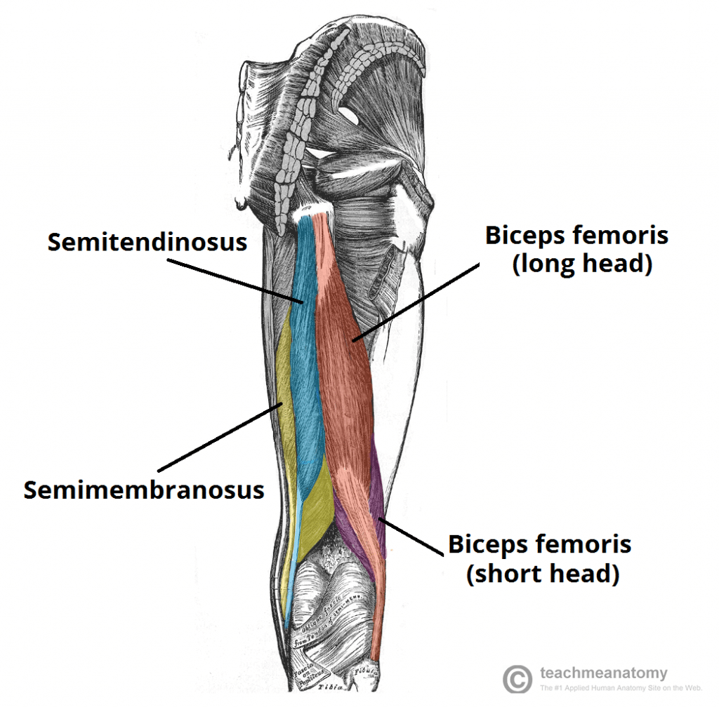
Muscles of the Posterior Thigh - Hamstrings - Damage ... from teachmeanatomy.info It adducts the thigh and flexes the leg at the knee. Jun 28, 2021 · the thigh is the region between the hip and knee joints. See full list on dummies.com The three layers of gluteal muscles, gluteus maximus, gluteus medius, gluteus minimus. The continuation of the quadriceps tendon that extends from the patella and inserts onto the tibial tuberosity of the tibia is called the patellar ligament. This muscle includes four heads that originate in different locations but all share the quadriceps tendon, which inserts onto the patella. This muscle originates on the pubis and the ischial tuberosity. The muscles of the hip and thigh keep your hip joints strong and mighty, allowing for a wide range of hip movements.
•medial thigh muscles•adductor longus muscle•adductor magnus muscle•adductor.
Vastus lateralis vastus medialis vastus intermedius rectus femoris This muscle includes four heads that originate in different locations but all share the quadriceps tendon, which inserts onto the patella. Its innervated by the obturator nerve and laterally rotates the thigh. It functions with the semitendinosus to extend the thigh and flex and medially rotate the leg. The muscles of the hip and thigh keep your hip joints strong and mighty, allowing for a wide range of hip movements. Jul 05, 2021 · upper limb anatomy arm anatomy muscle anatomy anatomy study body anatomy anatomy thigh: Human muscle anatomy 12 photos of the human muscle anatomy human anatomy muscle questions, human anatomy muscles clay learning system, human muscle anatomy head, human muscle anatomy leg, human muscle anatomy worksheet, human muscles, human anatomy muscle questions, human anatomy muscles clay learning system, human muscle. Like the forearm, the upper leg, or thigh, has a dense arrangement of many muscles. This muscle originates on the superior ramus of the pubis portion of the hip bone and inserts on the pectineal line of the femur. It's also the largest joint in the body. Its innervated by the obturator nerve. Originating at the obturator foramen and membrane of the hip bone, this muscle inserts onto the femur. Anatomy books made for artists!
Ebraheim's educational animated video describes muscle anatomy of the thigh. On the anterior side, the most prominent of the muscles are the sartorius muscle and the four muscles that make up quadriceps muscle group (the "quads".) 2, vastus medialis & intermedius muscles. Originating on the pubis and inserting on the pectineal line and linea aspera of the femur, this muscle is innervated by the obturator nerve. It also flexes the leg at the knee.
Upper Thigh Anatomy - Muscle compartments of the Thigh ... from www.wesnorman.com The knee joins the upper leg and the lower leg. The most medial of the three hamstring muscles, this muscle originates on the ischial tuberosity and inserts on the medial condyle of the tibia. The rectus femoris, however, also flexes the hip. Anatomy books made for artists! It adducts the thigh and flexes the leg at the knee. The single bone in the thigh is called the femur. This muscle originates on the superior ramus of the pubis portion of the hip bone and inserts on the pectineal line of the femur. 2, vastus medialis & intermedius muscles.
Jun 18, 2018 · leg anatomy upper leg anatomy and function.
Its innervated by the tibial portion of the sciatic nerve. Jul 20, 2016 · related posts of muscle anatomy of upper thigh human muscle anatomy. Like the forearm, the upper leg, or thigh, has a dense arrangement of many muscles. Its innervated by the obturator nerve. Jun 18, 2018 · leg anatomy upper leg anatomy and function. See full list on dummies.com Vastus lateralis vastus medialis vastus intermedius rectus femoris Ebraheim's educational animated video describes muscle anatomy of the thigh. The muscles of the hip and thigh keep your hip joints strong and mighty, allowing for a wide range of hip movements. See full list on dummies.com More images for upper thigh anatomy » It's also the largest joint in the body. 2, vastus medialis & intermedius muscles.